Making a clinical neurological assessment impossible.Īn MRI was performed prior to surgical stabilisation, 19 shows a distracted type 2/3 odontoid peg injury in a patient who also sustained a serious head injury, It may also be useful in problem solving to distinguish between acute and old fractures.įig. MRI is useful in assessing soft tissue and neurological injury. Īlthough CT is the gold standard for bony anatomical detail, This is thought to occur secondary to a previous fracture of the synchondrosis and may cause instability.Ī persistent ossiculum terminale is due to failure of fusion of the secondary ossification centre,Ĭloser to the tip of the odontoid process than the synchondrosis,Īnd may mimic a type 1 fracture ( Fig. One should be aware of anatomical variations which may mimic fractures. Prompting further investigation with MRI.

17 demonstrates a haematoma in the spinal canal, Haematoma may point to a subtle fracture in a degenerative neck or disruption of the posterior ligamentous complex.įig. Useful information may also be gained by reviewing the CT in the soft tissue window. The radiologist should also remember to review this area in 3 orthogonal planes on CT head examinations,Īs peg fractures may not be clinically suspected in elderly patients who have had a fall ( Fig. They may not be apparent on axial imaging,Īnd hence coronal and sagittal reformats should be obtained. It is also useful to demonstrate the anatomy of fractures in order to aid surgical planning.ĭue to the transverse plane of odontoid fractures,

14).ĬT is essential to rule out minimally displaced fractures in patients with inadequate radiographs or degenerative disease. It may not be possible to exclude fractures on plain radiographs when there are degenerative changes present,Įven on CT degenerative changes can draw one’s eye away from acute fractures ( Fig.

They may be clearly seen on an open mouth view ( Fig. Or demonstrate prevertebral soft tissue swelling ( Fig. 11). Lateral radiographs may appear normal in the case of undisplaced Type 2 or 3 fractures, 10).Ī high degree of suspicion for peg fractures is needed when reviewing imaging of the head and cervical spine.ĭisplaced odontoid peg fractures can be identified on lateral radiographs ( Fig. One should ensure careful examination in 3 orthogonal planes to exclude extension of the fracture into the posterior elements ( Fig. Īs they involve the body of the axis and have a relatively large surface area they are relatively stable and have a much lower risk of malunion ( Fig. Making it more unstable and definitely necessitating surgical stabilisation to prevent non-union ( Fig. Īlso involves additional chip fracture fragments at the base of the dens, Hadley et al suggested a modification of the Anderson & D’Alonzo classification system with the inclusion of a subtype of type 2 odontoid process fractures (type 2A). The risk of non-union is directly related to the degree of displacement ( Fig. Probably due to disruption of blood supply to the peg [5, With a risk of atlantoaxial subluxation if not promptly immobilised ( Fig.

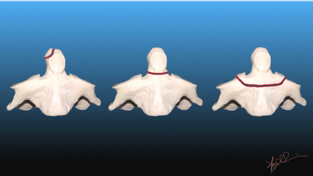
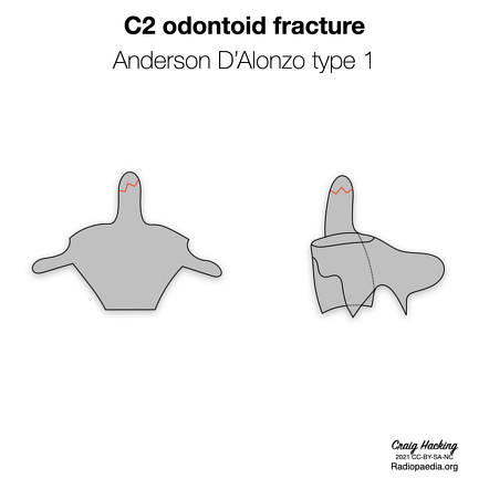
Or an associated occipital condyle fracture. They may be unstable if there is bilateral alar ligament avulsion, They are thought to be avulsion fractures of the alar or apical ligaments and occur above the level of the transverse ligaments, Type 1 odontoid process fractures are rare and somewhat controversial. Oblique fracture through upper part of the odontoid process caused by bony avulsion by alar ligamentsįracture at junction between the odontoid process and the body of the axisįracture that extends down into the cancellous bone of the body of the axis and is in reality a fracture of the body of C2 The fracture types may then be further classified as displaced or non-displaced. The classification system is summarised in Table 1 and Fig. They characterised odontoid peg fractures according the plane of the fracture. The Anderson and D’Alonzo classification system is the most widely used. Neurological injury is uncommon but due to the risk of atlantoaxial subluxation and proximity of the medulla oblongata, Odontoid fractures are the most common isolated cervical spine fracture in patients over the age of 70 years. Such as road traffic collisions in young or middle-aged patients. Odontoid peg fractures are thought to occur following extreme hyperextension or hyperflexion of the cervical spine.


 0 kommentar(er)
